Morphological Features of the Testes of the Liver Fluke, Opisthorchis viverrini
Abstract
Background: Infection of Opisthorchis viverrini is widely endemic mainly in Northeast of Thailand including Laos, Cambodia and South Vietnam. Its prevalence and pathogenic effects are associated with several hepatobiliary diseases in human. The liver fluke is monoecious, seft - fertilization can be occurred individually. The male reproductive organ usually consists of two deeply lobed testes, located in the posterior region of the body next to the ovary. No description is available on the ultrastructure and development of the testes of the Opisthorchis viverrini.
Objectives: This study aims to examine the morphology and ultrastructure of the testes of the adult flukes O. viverrini.
Study design: Descriptive study based on morphological observation by using light microscope (LM), transmission electron microscope (TEM) and scanning electron microscope (SEM).
Setting: Department of Anatomy and Department of Parasitology, Faculty of Medicine, Khon Kaen University, Thailand.
Materials and Methods: The encyst metacercaria of Opisthorchis viverrini were identified and collected. Ten adult Golden Syrian hamsters were used. Each animal was infected with 50 encyst metacercaria by gastric intubation. The animal were then sacrificed by deep ether anaesthesia, at 28 days post infection. The recovered flukes were routinely processed for investigation of the testes by LM, TEM and SEM.
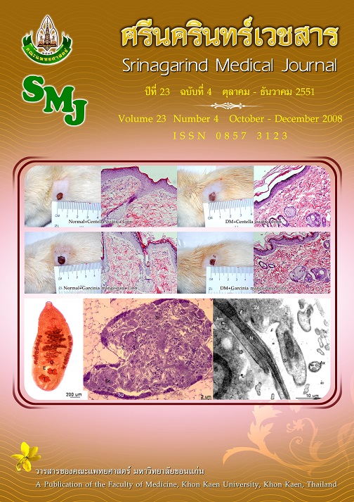
Results: In the whole mount specimens, the pair testes of the adult O. viverrini were clearly defined at the posterior 1/3 of the worm body posterior to the ovary. They were multilobate, 4-5 lobes each and location one behind the other. The testes showed numerous germ cells at various stages of development. The spermatogonia were closest to the testicular wall. In the deeper regions, cluster of numerous spermatocytes and spermatids were observed. The early spermatid exhibited an elongated nucleus with exhibiting lamellar chromatin alongside the two sets of striated rootlets. The scanning electron microscopic studied revealed that the
spermatozoa were long filiform-shaped.
Conclusions: This is the first report of the morphology of the testes of the adult flukes, Opisthorchis viverrini studied by using light and electron microscopes. The testes contained different stages of developing spermatogenic cells. Their three dimentional morphology were thread-like with no easily discernible heads and tails.
Keywords: Opisthorchis viverrini, spermatogenic cells, testes, ultrastructure