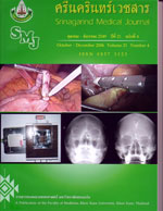
Surgical Management of Laryngo-tracheo-esophageal Cleft : the First 2 Cases Experience at Srinagarind Hospital
Abstract
Background : Laryngo-tracheo-esophageal cleft (LTEC) is a rare congenital anomaly resulting from failure of the posterior cricoid larmina to fuse and incomplete development of tracheoesophageal septum . In symptomatic cases, early presentation is with feeding problems, choking, chronic coughing, wheezing, cyanotic spells and apnea. Aspiration pneumonia may result and cause of life-threatened morbidity. The awareness of unusual clinical features of LTEC will lead to early investigations and diagnosis, which can prevent further complications. Surgery is the treatment of choice to correct this anomaly. These 2 LTEC cases would make medical personals familiar with clinical presentation and surgical management.
Objective : To review and report surgical management in 2 cases of LTEC at Srinagarind Hospital
Study Design : Case report by clinical data collection from medical record of LTEC patients who admitted during 1 December 2005 and 31 July 2006
Results : These 2 LTEC cases recently presented within 5 month-interval at Srinagarind Hospital throughout the last 20 years. Both cases had the same symptoms of dyspnea, cyanosis and aspiration pneumonia. Only one case is still alive after definite surgery. The first LTEC was a 1800-gram-male newborn of 36 year-old mother, G1P0, GA 42 wks. He was normally labored with APGAR score 8,9,10, and developed dyspnea and cyanosis after 1 hour later. His anomaly was detected during intubation and needed 2 endotracheal tubes insertion. Under direct laryngoscopic and bronchoscopic finding, the LTEC classified as type VI (complete cleft from arythenoid throughout carina). He had severe aspiration pneumonia and needed gastrostomy tube for decompression. And he finally got esophageal ligation to completely control regurgitation and jejunostomy tube for enteral feeding. The definite surgery was planed as soon as his weight reached 3 kg. Unfortunately, he got severe pneumonia and septicemia, and terminated with respiratory failure.
The second case was a 3000 gm female newborn of 19 year-old mother,G1P0, GA 39 wks. She was obviously diagnosed anal atresia with ano-vestibular fistula, and later suspected LTEC due to frequent endotracheal tube dislodgement. Direct laryngoscopy and bronchoscopy identified LTEC type III (cleft extent from arythenoids to distal trachea at level of 2.5 cm above carina). She got gastrostomy tube for gastric decompression and jejunostomy tube for early feeding. Finally the surgical separation of trachea and esophagus was performed on the 33rd day of live. She has had excessive secretion and needed tracheostomy tube for secretion clearance. At present, she can suck nibble well but hardly swallow infant formular. The radiological investigations reviewed slightly narrowing of the esophageal lumen without leakage. The further surgical plan is esophageal dilatation and correction the rest laryngeal cleft. The development of laryngeal function should be periodic examined and physiotherapy are considered to improve air way clearance.
Conclusions : Even LTEC is a rare aerodigestive anomalies, intensive medical care is needed throughout the admission. Early diagnosis is the important factor to decrease risk of complications. Surgery is the main treatment for survival. Postopertive care is necessary to improve the quality of life.
Key words : laryngo-tracheo-esophageal cleft, laryngotracheal cleft, posterior laryngeal cleft


