Association between Urinary Tract Infection, Gender, Age and the Occurrence of Magnesium Ammonium Phosphate compared with Calcium Oxalate Uroliths in Dogs
Main Article Content
Abstract
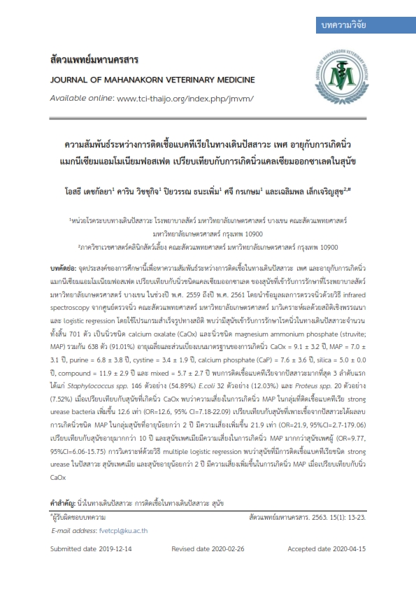
The objectives of this study were evaluated the associations between urinary tract infection, gender, age and magnesium ammonium phosphate urolith occurrences compared with calcium oxalate urolith occurrences in dogs visited at Kasetsart University Veterinary Teaching Hospital during 2016-2018. Urolith compositions were analyzed at Kasetsart University Urolith Center by infrared spectroscopy. Descriptive statistic and logistic regression were performed by the commercial software. The results of 701 urolith samples were analyzed. The common uroliths (638 submitted samples; 91.01%) were calcium oxalate (CaOx) and magnesium ammonium phosphate (struvite; MAP). The average age and standard deviation for dogs with CaOx uroliths was 9.1 ± 3.2 years, 7.0 ± 3.1 years for MAP, 6.8 ± 3.8 years for Purine, 3.4 ± 1.9 years for cystine, 7.6 ± 3.6 years for calcium phosphate (CaP), 5.0 ± 0.0 years for silica, 11.9 ± 2.9 years for compound and 5.7 ± 2.7 years for mixed uroliths. The top 3 bacterial urinary tract infections were 146 (54.89%) Staphylococcus spp., 32 (12.03%) E.coli, and 20 (7.52%) Proteus spp. Comparison with dogs with CaOx uroliths, the dogs with strong urease bacterial urinary tract infection has increased risk (OR=12.6, 95% CI=7.18-22.09) for developing MAP uroliths compared with dogs with no growth urine sample. The dogs less than 2 years old had increased risk (OR=21.9, 95%CI=2.7-179.06) for developing MAP uroliths compared with over 10 years old dogs. Female dogs had increased risk (OR=9.77, 95%CI=6.06-15.75) for developing MAP uroliths compared with male dogs. The result of multiple logistic regression indicated that dogs with strong urease bacterial urinary tract infection, female dogs and dogs less than 2 years old had increased risk for developing MAP uroliths compared with dogs with CaOx uroliths.
Article Details
References
Adsanychan N., S. Hoisang, S. Seesupa, N. Kampa, P. Kunkitti, and S. Jitpean. 2019. Bacterial isolates and antimicrobial susceptibility in dogs with urinary tract infection in Thailand: a retrospective study between 2013-2017. Vet. Integr. Sci. 17(1): 21-31
Detkalaya O., S. N. Detkalaya and C Lekcharoensuk. 2017. Epidemiology of Canine Urolithiasis in Thailand during 2006-2013. J. Kasetsart Vet. 27: 38-53.
Hanhaboon P., C. Bumrungpun, S. Wongnarkpet and P. Amavisit. 2011. Occurrences and Antimicrobial Resistant Rates of Bacteria isolated from Dog Patients of KU Veterinary Teaching Hospital in 2008. J. Thai Vet. Pract. 24 (3-4): 15-21.
Lekcharoensuk, C., C. A. Osborne, J. P. Lulich, R. Pusoonthornthum, C. A. Kirk, L. K. Ulrich, L. A. Koehler, K. A. Carpenter, and L. L. Swanson. 2002. Associations between dietary factors in canned food and formation of calcium oxalate uroliths in dogs. Am. J. Vet. Res. 63(2): 163-169.
Lulich, J. P., C. A. Osborne, H. Albasan, L. A. Koehler, L. M. Ulrich, and C. Lekcharoensuk. 2013. Recent shifts in the global proportions of canine uroliths. Vet. Rec. 172(14): 363.
Lulich J. P., A. C. Berent, L. G. Adams, J. L. Westropp, J. W. Bartges, and C. A. Osborne. 2016. ACVIM small animal consensus recommendations on the treatment and prevention of uroliths in dogs and cats. J. Vet. Intern. Med. 30: 1564–1574.
Ling, G. V., C. E. Franti, A. L. Ruby, and D. L. Johnson. 1998. Urolithiasis in dogs. Ii: breed prevalence, and interrelations of breed, sex, age, and mineral composition. Am. J. Vet. Res. 59(5): 630-642.
Osborne, C. A., J. P. Lulich, J. M. Kruger, L. K. Ulrich, and L. A. Koehler. 2008. Analysis of 451,891 canine uroliths, feline uroliths, and feline urethral plugs from 1981 to 2007: Perspectives from the Minnesota Urolith Center. Vet. Clin. North Am. Small Anim. Pract. 39(1): 183-197.
Rebecca S. and J. W. Bartges. 2001. Canine struvite urolithiasis. 23(5): 407-420.
Roe, K., A. Pratt, J. Lulich, C. Osborne, and H. M. Syme. 2012. Analysis of 14,008 uroliths from dogs in the UK over a 10-year period. J. Small Anim. Pract. 53(11): 634-640.
Wong C., S. E. Epstein, and J. L. Westropp. 2015. Antimicrobial susceptibility patterns in urinary tract infections in dogs (2010–2013). J. Vet. Intern. Med. 29: 1045–1052.


