Effects of Operating Modes and Electric Potential Difference on Image Quality and Elemental Analysis of Zinc Oxide Nanomaterials
Article Sidebar
Main Article Content
Abstract
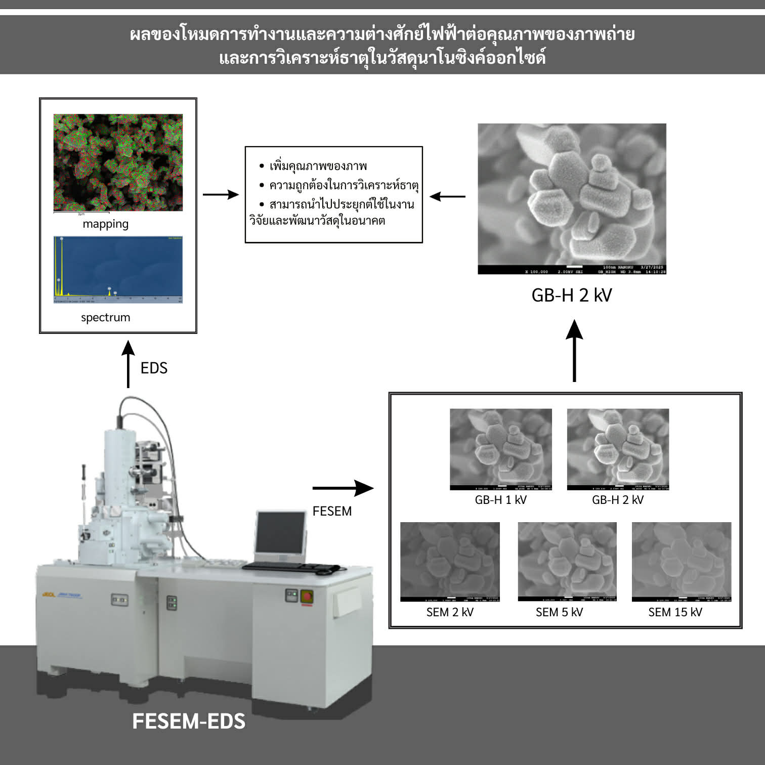
This research focuses on selecting the optimal operating mode and accelerating voltage for the analysis of zinc oxide (ZnO) nanomaterials using a Field Emission Scanning Electron Microscope (FESEM) combined with Energy Dispersive X-ray Spectroscopy (EDS). The effects of operating mode and accelerating voltage on image quality and elemental analysis were evaluated. The results showed that the GB-HIGH mode at an accelerating voltage of 2 kV produced the sharpest images and revealed the most detailed surface morphology compared with the SEM mode and other accelerating voltages. In addition, EDS analysis indicated a uniform distribution of zinc (Zn) and oxygen (O) on the material surface. A comparison between the Analyzer method and the Point & ID method for elemental quantification revealed similar Zn and O contents, consistent with the characteristics of ZnO nanomaterials. These results highlight the importance of selecting appropriate operating parameters to enhance image quality and improve the accuracy of elemental analysis in nanomaterial research, and they can be applied to future materials research and development.
Article Details

This work is licensed under a Creative Commons Attribution-NonCommercial-NoDerivatives 4.0 International License.
Authors retain the copyright of articles published in Wichcha Journal Nakhon Si Thammarat Rajabhat University. All published articles are distributed under the Creative Commons Attribution–NonCommercial–NoDerivatives License (CC BY-NC-ND 4.0) (https://creativecommons.org/licenses/by-nc-nd/4.0/). Under this license, readers are permitted to read, download, and share the articles for non-commercial purposes, provided that proper attribution to the original source is given and the content is not modified or altered. All contents of the articles, including text, tables, figures, equations, and other illustrations, are the sole responsibility of the authors. The views and opinions expressed in the articles do not necessarily reflect those of the editorial board or the publisher.
References
ชีวะ ทัศนา นิคม ผึ่งคำ สมยศ ศรีคงรักษ์ จันทนีย์ เพ็ชรไพบูลย์ และวิลาสินี เนินริมหนอง. (2561). ผลของอนุภาคนาโนซิงค์ออกไซด์ต่อการเจริญเติบโตของพริกหวาน. วารสารมหาวิทยาลัยทักษิณ, 21(3), 51-57.
ดลฤดี โตเย็น. (2562). การวิเคราะห์ธาตุและองค์ประกอบด้วยเทคนิค energy dispersive x-ray spectroscopy (EDS). สืบค้นเมื่อ 15 กันยายน 2567, จาก: https://www3.rdi.ku.ac.th/cl/knowledge/2562/EDS.pdf.
ดลฤดี โตเย็น. (2563). การเตรียมตัวอย่างทางวัสดุศาสตร์สำหรับการวิเคราะห์ด้วยกล้องจุลทรรศน์อิเล็กตรอนแบบส่องกราด. สืบค้นเมื่อ 15 กันยายน 2567, จาก: https://www3.rdi.ku.ac.th/cl/knowledge/2563/material_prep.pdf.
ยุพดี เผ่าพันธ์. (2567). ประเด็นปัญหาที่พบบ่อยในการบันทึกภาพด้วยกล้องจุลทรรศน์อิเล็กตรอนแบบส่องกราด. สืบค้นเมื่อ 20 ตุลาคม 2567, จาก: https://www3.rdi.ku.ac.th/cl/knowledge/2567/jun/problemfromsem_savepic.pdf.
Ahmed, T., Wu, Z., Jiang, H., Luo, J., Noman, M., Shahid, M., Manzoor, I., Allemailem, K.S., Alrumaihi, F. and Li, B. (2021). Bioinspired green synthesis of zinc oxide nanoparticles from a native Bacillus cereus strain RNT6: Characterization and antibacterial activity against rice panicle blight pathogens Burkholderia glumae and B. gladioli. Nanomaterial, 11(4), 884, doi: https://doi.org/10.3390/nano11040884.
Chaisorn, W., Nuengmatcha, P., Noypha, A., Pimsen, R., Porrawatkul, P., Kuyyogsuy, A., Thepchuay, Y., Sricharoen, P., Limchoowong, N., Chanthai, S. and Nuengmatcha, P. (2023). Adsorption-photocatalytic degradation abilities of γ-irradiated chitosan-ZnO-AgNP composite for organic dye removal and antibacterial activity. Environmental Science and Pollution Research, 30, 96840-96859, doi: https://doi.org/10.1007/s11356-023-29305-y.
Demissie, M.G., Sabir, F.K., Edossa, G.D. and Gonfa, B.A. (2020). Synthesis of zinc oxide nanoparticles using leaf extract of Lippia adoensis (Koseret) and evaluation of its antibacterial activity. Journal of Chemistry, 2020, 7459042, doi: https://doi.org/10.1155/2020/7459042.
Habibi, M.H. and Karimi, B. (2014). Application of impregnation combustion method for fabrication of nanostructure CuO/ZnO composite oxide: XRD, FESEM, DRS, and FTIR study. Journal of Industrial and Engineering Chemistry, 20(4), 1566-1570, doi: https://doi.org/10.1016/j.jiec.2013.07.048.
JEOL Ltd. (2012). JSM-7600F field emission scanning electron microscope operation guide (Version ISM7600F-OG-1b). Tokyo: JEOL Ltd.
Kumar, S.S., Venkateswarlu, P., Rao, V.R. and Rao, G.N. (2013). Synthesis, characterization and optical properties of zinc oxide nanoparticles. International Nano Letters, 3(1), 30, doi: https://doi.org/10.1186/2228-5326-3-30.
Mandal, A.K., Katuwal, S., Tettey, F., Gupta, A., Bhattarai, S., Jaisi, S., Bhandari, D.P., Shah, A.K., Bhattarai, N. and Parajuli, N. (2022). Current research on zinc oxide nanoparticles: Synthesis, characterization, and biomedical application. Nanomaterial, 12(17), 3066, doi: https://doi.org/10.3390/nano12173066.
Newbury, D.E. and Ritchie, N.W.M. (2013). Is scanning electron microscopy/energy dispersive x-ray spectrometry (SEM/EDS) quantitative?. Scanning, 35, 141-168, doi: https://doi.org/10.1002/sca.21041.
Porrawatkul, P., Pimsen, R., Kuyyogsuy, A., Rattanaburi, P. and Nuengmatcha, P. (2024). Morphology-dependent photocatalytic performance of ZnO nanostructures in organic dye and antibiotic degradation. International Journal of Environmental Science and Technology, 21, 7397-7414, doi: https://doi.org/10.1007/s13762-024-05530-x.
Porrawatkul, P., Rattanaburi, P., Nuengmatcha, P., Kuyyogsuy, A. and Pimsen, R. (2023). Effect of Na and Al doping on ZnO nanoparticles for potential application in sunscreens. Journal of Photochemistry and Photobiology, B: Biology, 240, 112668, doi: https://doi.org/10.1016/j.jphotobiol.2023.112668.
Stevens, S.M., Jansson, K., Xiao, C., Asahina, S., Klingstedt, M., Grüner, D., Sakamoto, Y., Miyasaka, K., Cubillas, P., Brent, R., Han, L., Che, S., Ryoo, R., Zhao, D., Anderson, M.W., Schuth, F. and Terasaki, O. (2009). An apprasial of high resolution scanning electron microscopy applied to porous material. JEOL News, 44(1), 17-22.
Syed, J. (2017). Scanning electron microscopy in oral research. Journal of the Pakistan Dental Association, 26(4), 189-195, doi: https://doi.org/10.25301/JPDA.264.189.
